
I have provided a response to an article written and published in Crisis Magazine which criticized some elements of the facts and findings of my investigation. (see https://reasontobelieve.com.au/exaggerations-and-eucharistic-miracles-but-not-in-buenos-aires-1996-comment-on-crisis-magazine-by-ron-tesoriero/)
There are some additional matters which I feel I need to present to give a better understanding of the details of my work.
1. Whilst there have been known incidents of a Eucharistic Miracle in 1992, 1994 and 1996 in the Santa Maria Church in Buenos Aires, it is only the 1996 case that I have reported on.
2. It was on 5 October 1999 that I went to the Santa Maria Parish Church in Buenos Aires and began my investigation. On that day I interviewed Padre Alexandro Pezet and Padre Eduardo Graham, priests from the parish, and Senora Emma Fernandez the Eucharistic Minister. I also filmed interviews of them. I have published their testimony as to what they say happened with the host that was found abandoned in that church on 18 August 1996. See pages 91 to 96 of my book My Human Heart.
3. In their testimony they emphatically stated that the glass tube containing a dark substance that was presented to me and Dr Castanon shown on pages 98 to 99 of my book My Human Heart, was the same dark substance (the communion host) that had transformed when placed in water on 18 August 1996. That dark substance that we were presented with in that glass tube is clearly seen in the photograph on page 97 of my book My Human Heart.
4. On 5 October 1999, a sample of the substance in the glass tube was taken for examination. The content from the glass tube was poured into a glass bowl. Dr Castanon severed a piece of the substance and placed it into a sterilised glass test tube along with some of the water that was in the original glass tube. That test tube was then labelled for identification. That test tube was then placed in a translucent plastic container and sealed. Dr Castanon and I then signed over the seal that had been placed.
I directed the procedure for the filming and the taking of the sample. I also filmed the procedure and filmed the taking of the sample.
I was not the only person who filmed the process. Also present was a representative of the Archdiocese of Buenos Aires who was engaged to film the process on behalf of the Archdiocese.
5. On 21 October 1999, Dr Castanon and I personally delivered the sample taken from the Buenos Aires 1996 transformation, to Forensic Analytical, a forensic laboratory in San Francisco for DNA investigation (the results I deal with at point #11 below). Forensic Analytical gave the case a file number (Case 199900441 – 1- 2).
After Forensic Analytical did their investigation, I directed that they send the sample to Dr Robert Lawrence of Delta Pathology in Stockton California. This they did on 2 March 2000 via Fedex under Tracking No. #811156269046.
6. Dr Robert Lawrence prepared a microscopic slide of the sample and then examined it. The sample, he said contained epidermis (outmost layer of skin) infiltrated with white blood cells. In his report he described “an aggregate of keratotic and parakeratotic debris with enmeshed leukocytes. Also, there are scattered minute aggregate of brown material comprised of septate hyphal fragments enmeshed in proteinaceous matrix”.
On 7 December 2000, Mike Willesee and I travelled to the United States to interview Dr Lawrence about his findings. Dr Lawrence had retained the microscopic slide of the sample in his possession pending our interview with him. Dr Lawrence was not told the source of the sample. During the interview Dr Lawrence placed the microscopic slide under his microscope and displayed for us on a monitor screen what he had focused on. The image from that screen I present as the last photo on page 99 of my book My Human Heart. Visible on that image is the tip of a ballpoint pen which Dr Lawrence used to point out the presence of white blood cells in the sample. His comments were “they were active living white cells at the time they were collected and there was some inflammatory process going on”. He was emphatic also in saying, “if this material had been placed directly into water after it had been taken off the body I would expect these cells to have dissolved within a few minutes to an hour or two.”
So, whilst Dr Lawrence had given his determination of the presence of the white blood cells, he saw those cells as being interconnected with the inflammation of tissue which he believed was epidermal tissue (skin).
7. Afterwards I proceeded to gain the opinion of other forensic pathologists as to what the material on the microscopic slide might be. The microscopic slide given to them was the same slide that was examined by Dr Lawrence. I engaged certain Australian scientists, Dr Peter Ellis, a Senior Lecturer in Forensic Medicine at Sydney University, and Dr Thomas Loy of Queensland University. Both supported the interpretation of Dr Lawrence: that the material appeared to be skin tissue. However, another scientist I engaged, Dr John Walker of Sydney University, had a different opinion. He said that the material looked like muscle tissue, not skin.
8. On 2 March 2001, I travelled with Mike Wilesse and Dr Castanon to Italy to seek the opinion of Professor Odoardo Linoli. He was involved in the investigation of the Eucharistic Miracle of Lanciano that happened in the 8th Century. He was of the opinion that it was muscle tissue, perhaps even heart muscle, but could not be sure.
I had a problem. Three scientists said it was skin. One said it was muscle. One said it may be heart. The same slides were being identified by different scientists as completely different tissue.
9. In an attempt to try and understand why there had been difficulty in the interpretation of the material in the sample by different pathologists, I decided to embark upon a dedicated program of self-education using the internet. For a whole year I gained access to university portals and undertook in-depth studies of forensic pathology and cell biology tutorials. In this exercise I realized that pathologists identify the type of tissue that they are looking at under the microscope by reading the tissues’ architecture. And that they use pathology atlases as a foundation reference tool. Comparing an unknown sample side by side with a verified standard “like with like” is a common core principle of diagnostic medicine.
I realised that by utilising the resources on the internet to examine digital histology atlases of both “epidermal tissue” and cardiac tissue I could get to understand the problem that scientists, that I had hitherto engaged, were encountering. I came to the conclusion that perhaps the Buenos Aires sample looked more like epidermal tissue than cardiac tissue.
10. However, this all changed on 18 August 2003. It happened in these circumstances. (I have to interpose here that I am a practicing Catholic and that I have in my own personal experience over a number of years become aware of the power of prayer and that God is an interventionist God. That He does answer prayers particularly when those prayers are intended to enhance His interest).
On 17 August 2003, I attended a 6pm Saturday Mass in my local parish. I said in prayer at that Mass, “Jesus please help me with the problems I am encountering in my investigations”. There were two problems in particular which I had.
i. The identification of some unusual cells in “blood” that had shed from a statue of Christ in Bolivia that I was examining, and
ii. The problem of the identification of the “tissue” (heart or epidermis) in the Eucharistic Miracle of Buenos Aires.
The next morning at 8am on 18 August 2003, I continued my research work using the internet. When I was discussing with Dr Walker the identification of material in the Bolivia statue case, he remarked that the unusual material in the sample looked like “the nuclei of liver cells”. I wanted to follow up on that lead.
At 8am on 18 August 2003, I typed in a Google search these words “nuclei of liver cells + histology + images”. On the University of Texas Medical Department Pathology Website, four photographs of microscopic images of human tissue came up.
Here are the images that were displayed.
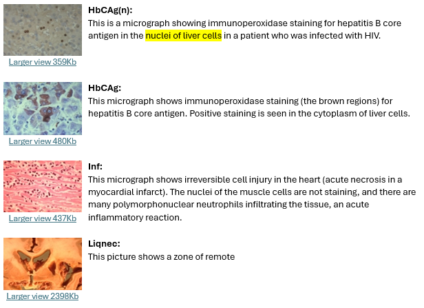
One showed the nuclei of liver cells. It was of no consequence. But the third image surprised me. It was an almost perfect copy of the photo taken from the microscopic slides of the Argentina communion host. The text accompanying it explained a lot. It read:
This micrograph shows irreversible cell injury in the heart (acute necrosis in a myocardial infarct). The nuclei of the muscle cells are not staining, and there are many polymorphonuclear neutrophils infiltrating this tissue; an acute inflammatory reaction.
I had keyed in words pertaining to the statue case and instead, by pure chance, had identified the tissue in the Argentina case.
This new information was alerting me to the fact that heart tissue changes in appearance when traumatised and it no longer looks like regular heart tissue. It occurred to me that the reason why the pathologists that I had previously engaged could not make an accurate identification was perhaps because they were not sufficiently familiar with the changes that progressively occur in the architecture of the heart tissue over intervals of time when there has been a compromising of the blood supply to the heart (myocardial infarction). (At a certain point in the progressive changes that occur the heart tissue can look more like what is typical of epidermis).
It is to be noted that in the Lanciano case that Professor Linoli was dealing with, the heart tissue did not show myocardial infarction. His uncertainty as to whether the material in the Buenos Aires sample was heart tissue or not could well have been because it did not have the regular appearance of heart tissue owing to changes that come about with myocardial infarction. Something that did not occur to him.
I realized that I needed to now consult an expert not only in forensic pathology, but in forensic cardiology.
I found that expert in New York, Dr Frederik Zugibe. He had a list of impressive qualifications which I set out in my book on page 105 of My Human Heart.
In my earlier posting on the internet in my article, I wrote about how I came to engage Dr Zugibe on the case.
I did not tell him anything about the history of the sample. In an email exchange, I simply said I was a lawyer working on a case and needed him to examine and interpret some material on a microscopic slide. I said that it was a confidential case, and I would tell him the history after he examined the sample.
On 20 April 2004, Australian journalist Mike Willesee and I travelled from Australia to New York to interview him. I brought with me and used a small professional video camera to record our interview with him. He placed the microscopic slide under his microscope and began to render his opinion. (The microscopic slide was the exact same one examined used by Dr Lawrence and the other previous scientific people I had consulted).
Dr Zugibe said that what he was looking at was heart muscle tissue from the left ventricle wall, not far from the valvular area. He then went on to say that this heart muscle is inflamed and lost its striations and is infiltrated with white blood cells. That the white blood cells are not normally found in heart tissue but are produced by the body and escape from the blood and infiltrate the heart tissue to address trauma and injury. There had been necrosis of the heart muscle. He also said that this was from a living person who had suffered traumatic injury where the blood supply to the heart had been compromised possibly by having been severely beaten across the chest. During the course of the interview I filmed Dr Zugibe go to a textbook on cardiology and show us the following:
A. That part of the heart, from where the heart muscle tissue had come and where the possible injury had occurred.
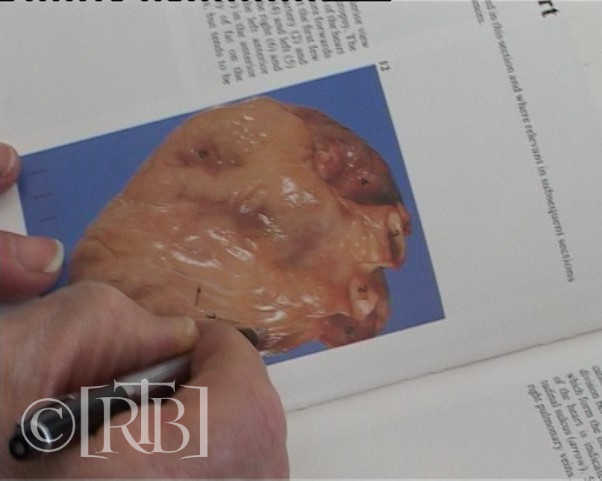
B. How he was able to identify what he had seen under the microscope was heart tissue undergoing degenerative changes. Noticeable in those displayed images was how the architecture of the tissue and cells change in time following myocardial infarction and how the subject case appears to be of a three-day old injury to the heart.
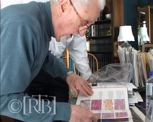
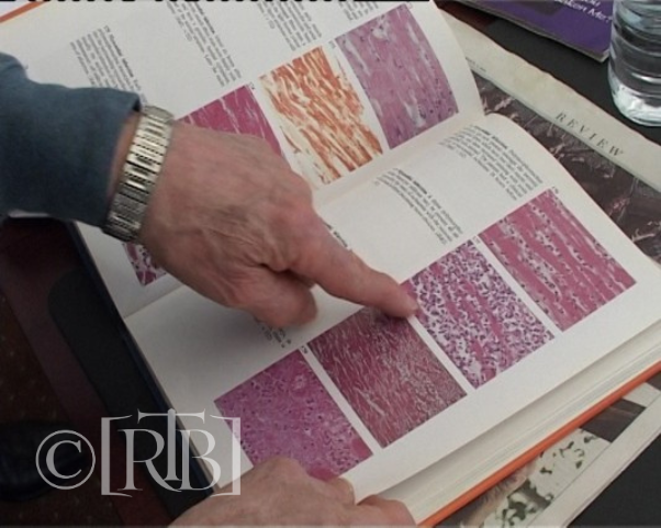
More of this interview can be read on pages 105 to 111 of my book My Human Heart and seen on my documentary which can be viewed here: https://www.youtube.com/watch?v=jimAT0bXhZM.
In our interview with Dr Zugibe he acquainted us with his expertise on forensic cardiology. He said that he is an expert on the heart and that the “heart” was his business.
As can be seen from his qualifications, Dr Zugibe is a most senior expert in his field of Forensic Cardiac Pathology. Amongst many of his qualifications are that he is the author of a leading textbook on Histopathology and was the Chief Medical Examiner of Rockland County of New York, Associate Professor of Pathology at Columbia University etc.
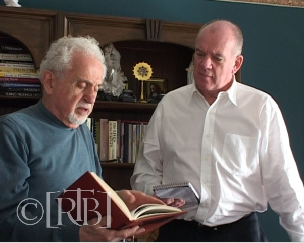
As such any report that he might issue on the examination of our case would carry the accuracy, gravity and reliability as we would expect from such a professional.
On 26 March 2005, Dr Zugibe sent to me his formal report of our meeting with him on 20 April 2004.
His report is most significant for the following reasons:
- He confirms that it was only Mike Willesee and I who attended his office on that date.
- That he examined the microscopic slide made, labelled and examined by Dr Lawrence of Delta Pathology.
- That I filmed and recorded Dr Zugibe as he rendered his opinion to Mike Willesee and myself.
- That he was not informed of the history of the material before he examined it.
- That he categorically identifies the material as cardiac muscle tissue infiltrated with inflammatory white blood cells.
- The myocardial infarction was of a few days duration.
Dr Zugibe’s clear and categorical statements above must surely dispel the thoughts of those who wish to express doubt, without evidence,:
- That Dr Zugibe did not know the origin of the material before he examined it.
- That he was exceptionally competent to determine that what he was looking at under the microscope was heart tissue, inflamed and human.
- That he provided a clear and unequivocal basis for his determination that it was inflamed heart tissue.
- That he intended to express clearly and without doubt everything that was contained in his formal report.
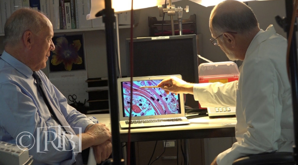
Subsequent to Dr Zugibe giving his formal report on 26 March 2005 on interpretation on the Buenos Aires case, other forensic pathologists have now confirmed the accuracy of Dr Zugibe’s interpretation. They include Dr Robert Lawrence who revised his opinion on 28 February 2008 at the meeting I had with him in San Francisco, and subsequently in an interview with Mike Willesee in September 2015 at the time the photograph below was taken. Also, confirmation of Dr Zugibe’s findings has recently been given by Italian cardiologist Dr Serafini.
Not long after I received Dr Zugibe’s report on the Buenos Aires case, I went back to retrace my research and to look again at the information that I had previously read on the University of Texas website on 18 August 2003. The micrograph that I saw on that website on that date gave this description of the image.

But, when I retraced my search on the item subsequently, the above image showed additional information which described the myocardial infarction of 3 to 4 days old.
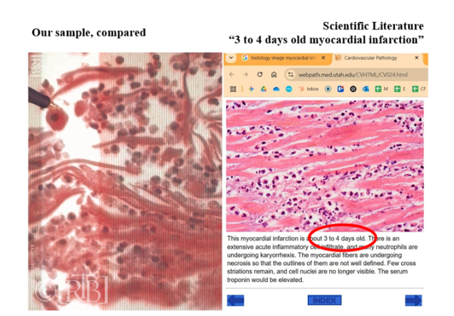
When you juxtapose the image of the Buenos Aires sample next to the scientific literature on the university website, one can see how Dr Zugibe could have come to his conclusion and that his interpretation was correct.
11. The issue of DNA studies on samples of the Eucharistic Miracle of Buenos Aires of 1996.
As stated previously, on 21 October 1999, Dr Castanon and I personally delivered to Forensic Analytical of San Francisco a sample for DNA analysis. They gave the case an identification number 9990441. The main object of the proposed testing was to see whether a human genetic profile could be obtained from the sample.
They sent to me their report of 1 May 2000. Significantly in that report they stated they were unable to generate a human genetic profile from the sample.
They summarized results of DNA PCR typing in the table as follows:
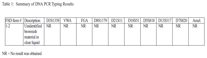
This result (or lack of result- if you like) has to be seen in the light of subsequent testing of the sample.
When Dr Lawrence of San Francisco looked at this sample under the microscope at our meeting with him on 7 December 2000, he remarked how the tissue was infiltrated with living white blood cells. He went on to say that those white blood cells were in a good state of preservation and that those cells would contain genetic material to identify the owner if DNA testing was done. Normally if there were white blood cells in a sample, DNA PCR testing should elicit the genetic profile of the owner of those blood cells. The fact that the testing did not show a genetic profile remains a mystery.
On 9 December 2005, I returned to Buenos Aires and obtained another sample from the 1996 Buenos Aires case. DNA testing on that sample was carried out in February 2016 using the latest in DNA testing technology involving the examination of single cells.
In a crime scene for example, when a sample is collected, blood cells may be mixed with skin cells and other contaminants. The laboratory we engaged to do DNA testing in 2016 had created breakthrough technology that could separate the cells from each other, select only the blood cells and then genetically analyze them with 100% accuracy. A number of white blood cells were identified and collected. Some were used in nuclear DNA testing and others in mitochondrial DNA testing (Details are set out in Chapter 27 of my book My Human Heart).
In nuclear DNA testing on white blood cells they were unable to obtain a human genetic profile. However, in mitochondrial DNA testing for maternal inheritance on single white blood cells revealed a surprising amount of genetic data. This was from the non-coding region of the human genome. They were able to obtain significant genetic information on the maternal ancestry of the person.
The genetic data obtained in these tests will be of immense value for comparative testing of blood stains on the Shroud of Turin in the future. In the meantime, the genetic data that we hold will remain confidential and will only be released at the time of the proposed future comparative testing with the Shroud of Turin.
To see my brief documentary presenting the case for the comparative testing with the Shroud of Turin, go to this link: https://youtu.be/jHa0INzcF-s?si=HKB4a3kXvl1Tadl0

